
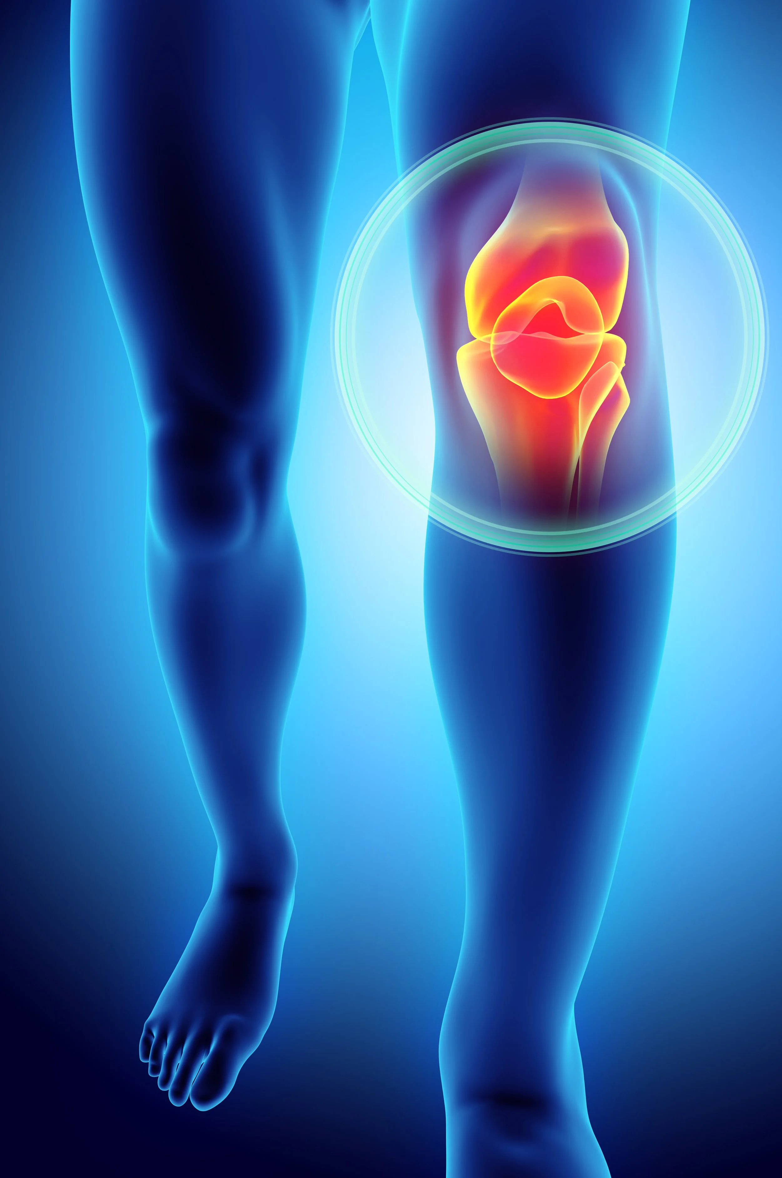
MPFL Reconstruction: How Dr. Raynor Stabilizes the Kneecap
When a kneecap keeps dislocating, the fix is a new ligament. MPFL reconstruction replaces the torn medial patellofemoral ligament with a tendon graft that runs from the inside edge of the kneecap to the inside of the thigh bone and catches the kneecap if it starts toward the outside. Dr. Brett Raynor performs it as an outpatient operation through two small incisions, usually with an arthroscopic look inside the knee first. Most patients walk on the leg the same day with the brace locked straight, are out of the brace at about six weeks, feel really good at three months, run at about four months, and return to cutting sports at about six months. When the shape of the knee is working against the kneecap, he may also move the bony attachment of the patellar tendon at the same time; that version takes longer.
This page covers the operation itself. For what a kneecap dislocation is and when surgery is the right call, see kneecap dislocations and patellar instability. For the week-by-week recovery, see the MPFL reconstruction recovery roadmap.
Before surgery
MPFL reconstruction is an elective operation, and timing is flexible. Because it is a reconstruction rather than a repair, the surgery is the same whether it is done a month or a year after the last dislocation. The one exception is a loose piece of cartilage in the joint, which needs to be dealt with promptly. For an athlete in season, that often means playing in a brace and scheduling the reconstruction for the off-season.
Before the operation Dr. Raynor reviews the MRI with each patient: the state of the cartilage on the back of the kneecap, the depth of the groove, and the TT-TG distance, which measures how far toward the outside of the shin the patellar tendon attaches. Those findings decide whether the new ligament is enough on its own or whether the tendon attachment should be moved as well. A knee with a tendon attachment 20 millimeters or more off center has anatomy that actively pulls the kneecap outward; that threshold comes from the original study that defined the measurement (Dejour et al., Knee Surg Sports Traumatol Arthrosc, 1994).
Surgery is done under general anesthesia with a nerve block in the thigh for pain control afterward. Patients go home the same day.
The operation, step by step
1. Examination under anesthesia. With the muscles fully relaxed, Dr. Raynor checks how far the kneecap can be pushed toward the outside and watches how it tracks as the knee bends. This is the clearest look at the instability he will get, and occasionally it changes the plan, for instance toward moving the tendon attachment in a knee that is looser than expected.
2. A look inside the knee. Through two small poke-hole incisions, an arthroscope lets him inspect the cartilage on the back of the kneecap and in the groove. Loose fragments are removed; a sizable piece of cartilage and bone that came off with a dislocation is fixed back in place when it can be. He also watches the kneecap track from inside the joint.
3. The graft. The new ligament is made from donor tendon, prepared on the back table while the knee is being examined. Using donor tissue means no second incision to harvest a tendon and no weakening of the patient's own hamstring or quadriceps, which matters when the quadriceps is the muscle that has to come back strongest afterward. In a review of 1,504 reconstructions, recurrent instability was no more common with donor tendon than with the patient's own tissue (McNeilan et al., Arthroscopy, 2018). The graft is folded so that it has two limbs on the kneecap side and one on the thigh-bone side, matching the fan shape of the native ligament.
4. Anchoring to the kneecap. Through a small incision along the inside edge of the kneecap, two anchors are placed into the bone at the inside edge, one toward the top and one toward the bottom. Each limb of the graft goes to its own anchor, the upper limb to the upper anchor and the lower limb to the lower anchor, without crossing them, so that the graft lies the way the original ligament did. The anchors sit in the edge of the bone rather than running tunnels across the kneecap, which keeps the kneecap strong.
5. Finding the attachment on the thigh bone. The MPFL attaches to a small saddle of bone on the inside of the thigh bone, between two bumps that can be felt through the skin. Getting this spot right is the single most important step of the operation: a graft attached a few millimeters too high or too low tightens or slackens as the knee bends, which leads either to a stiff, painful knee or to a kneecap that can still come out. Dr. Raynor locates the point by feel and confirms it with a live X-ray (fluoroscopy) in the operating room against a published landmark (Schöttle et al., Am J Sports Med, 2007); he does this on every case. A small socket is then drilled at that point. The angle of the socket depends on the patient. When the growth plates are closed, it is angled upward and forward, away from the back of the knee. When the growth plates are still open, it is angled downward, away from the growth plate, so the growth of the leg is not affected.
6. Passing and tensioning the graft. The single limb of the graft is passed under the tissue layer on the inside of the knee from the kneecap incision to the thigh-bone incision. Tension is the step that takes judgment. With the knee bent to about 30 degrees, the point at which the kneecap has settled into its groove, the graft is set so that the kneecap sits centered and the ligament is just snug: tight enough that the kneecap cannot slide over the edge of the groove, loose enough that the kneecap still moves a little side to side the way a normal kneecap does. Dr. Raynor then runs the knee through its full motion to check that the graft stays at the same tension from straight to fully bent, and fixes the graft in the socket with a screw that wedges it against the bone (an interference screw). When the growth plates are closed, the graft is pulled into the socket and the screw is placed beside it; when they are open, the graft and screw are pushed into the socket together, which keeps everything below the growth plate.
7. Closing. The incisions are closed with dissolvable stitches under the skin and skin glue, the knee is dressed, and a brace is locked straight for walking. The whole operation usually takes about an hour to an hour and a half, and Dr. Raynor takes as long as it needs to get the tension and the attachment point exactly right.
When the tendon attachment is moved as well
In some knees the patellar tendon attaches far enough toward the outside of the shin that the pull of the thigh muscle angles the kneecap outward with every step. A new ligament alone is working against that angle, and in a knee that has dislocated repeatedly it can fail. In those patients Dr. Raynor also moves the bony attachment of the patellar tendon (a tibial tubercle osteotomy). Through an incision on the front of the shin, he cuts the bump of bone the tendon attaches to, with the cut angled so that it comes back around the bump rather than running straight across the shin, slides the bump toward the inside and slightly forward, and fixes it in its new position with screws. Shifting it inward straightens the line of pull; shifting it forward takes pressure off the cartilage on the back of the kneecap, which is why this operation was first described for painful, worn kneecaps (Fulkerson et al., Am J Sports Med, 1990).
The moved bone has to heal like a fracture, so this version of the operation changes the recovery: no weight on the leg for six weeks, the brace on for walking and sleeping during that time, and motion opened up in steps. The whole roadmap runs two to three months longer. Dr. Raynor does not add the bone work routinely. He reserves it for knees that have dislocated more than once and have the measurements to justify it, because for an adult who is not dislocating all the time, it would be an aggressive first operation.
How often does it hold?
MPFL reconstruction is one of the most reliable operations in sports medicine. In a review that pooled 14 studies of isolated MPFL reconstruction, the kneecap dislocated again in 1.2 percent of patients, and 84 percent returned to sport (Schneider et al., Am J Sports Med, 2016). A review of 629 knees that also counted partial slips found recurrent instability in 3.7 percent (Shah et al., Am J Sports Med, 2012). For comparison, the older approach of tightening the torn tissue rather than replacing it failed in about 1 in 3 knees (Camp et al., Am J Sports Med, 2010), which is why Dr. Raynor reconstructs rather than repairs.
Those numbers describe the kneecap staying in. The same 629-knee review found that about 1 in 4 patients had some complication after surgery, most commonly stiffness, persistent pain along the inside of the knee, or a lingering sense of apprehension about the kneecap, with true redislocation being rare. Most of those problems are treated with therapy. Dr. Raynor's patients are told that plainly: the operation is excellent at its main job, and getting the knee moving early is how the stiffness is avoided.
Risks
Every operation has risks. The ones specific to MPFL reconstruction are:
Stiffness, the most common problem, which is why motion starts right away in therapy
Pain on the inside of the knee, usually from a graft that is slightly too tight, and more common in adults than in teenagers; it often settles with time and therapy, and rarely needs the graft loosened
Another dislocation, in about 1 to 4 of every 100 patients
Fracture of the kneecap, rare with edge anchors, but the reason tunnels through the kneecap are avoided
Growth plate injury in a growing patient, which the physeal-sparing socket is designed to prevent
Infection, blood clots and numbness around the incisions, as with any knee surgery; a baby aspirin a day for about six weeks lowers the clot risk
Recovery
The full stage-by-stage plan is on the MPFL reconstruction recovery roadmap, and the MPFL reconstruction FAQ answers the practical questions about showering, driving and work. In brief:
Surgery to week 2. Home the same day. Full weight on the leg with the brace locked straight; the crutches are for balance. With the knee straight the kneecap sits above its groove, where it is much less likely to dislocate. Bending to 90 degrees in therapy, which starts within days.
Weeks 2 to 6. The brace unlocks once the quadriceps can hold the leg straight, usually at two to three weeks; crutches go in the same window. Brace off at about six weeks. Desk work and school in the first week or two.
Weeks 6 to 12. Feeling good. Walking normally, stationary bike, elliptical, light weights. Driving once off crutches and off prescription pain medication, usually about six to eight weeks for a right knee.
Months 3 to 6. Really good at three months. Running at about four months, once the first part of the sport test is passed.
About 6 months. Cutting and pivoting sports after passing a sport test with the physical therapist, with the quadriceps at 90 percent or better of the other leg.
When the tendon attachment is moved as well, the first six weeks are on crutches without weight on the leg, and running and sport come closer to four to six months and eight to nine months.
Why patients choose Dr. Raynor for MPFL reconstruction
Dr. Raynor treats kneecap instability across its whole range, from the high school athlete after a first dislocation to the adult whose kneecap has been loose for years, and he is deliberately conservative about who needs an operation. When surgery is the right call, MPFL reconstruction has been one of his most dependable operations across hundreds of cases, and he adds the bone work only when the anatomy requires it. He is the team physician for the U.S. Ski and Snowboard Team, Highland Park High School, St. Mark's School of Texas, Ursuline Academy and Hockaday, trained at the Steadman Philippon Research Institute in Vail after his orthopaedic residency at Vanderbilt, and has been named a D Magazine Best Doctor every year since 2019.
-
Yes. Patients go home the same day with a nerve block in the thigh, numbing medicine in the incisions, and a prescription for pain medication, and most need the stronger medication only for the first few days. Ibuprofen and Tylenol do most of the work after that. A baby aspirin a day for about six weeks lowers the risk of a blood clot.
-
Using the patient's own hamstring or quadriceps tendon means a second incision, a harvest site that hurts, and a weaker muscle in a leg that needs its quadriceps back as fast as possible. Donor tendon avoids all of that, and the research does not show a higher rate of the kneecap coming out again with donor tissue (McNeilan et al., Arthroscopy, 2018). Donor tendon is screened, processed and sterilized, and the body incorporates it over the months after surgery.
-
When the MRI measurement of the tendon attachment (the TT-TG distance) is about 20 millimeters or more, the kneecap has dislocated more than once, and especially when the cartilage behind the kneecap is already worn. In those knees a new ligament alone is fighting the angle of pull, and moving the attachment inward and forward both straightens the pull and unloads the cartilage. It is a bigger operation with six weeks on crutches, so Dr. Raynor reserves it for knees that have earned it.
-
Yes. The growth plate at the end of the thigh bone sits close to where the MPFL attaches. In a growing patient, Dr. Raynor places the femoral socket just below the growth plate and angles it away, so the ligament can be rebuilt without disturbing growth. The recovery roadmap is the same. The sport test at the end matters even more in young athletes, whose quadriceps shuts down hard after a swollen knee and comes back slowly.
-
If a piece of cartilage and bone came off the kneecap or the thigh bone, it is dealt with at the same operation: fixed back in place when it is large enough and the bone on it will heal, removed when it is not. Cartilage work can add weight-bearing or motion limits in the first six weeks, and the post-op instructions spell those out.
-
Rarely. Across the published studies, about 1 to 4 of every 100 patients have another dislocation or a partial slip after MPFL reconstruction (Schneider et al., Am J Sports Med, 2016; Shah et al., Am J Sports Med, 2012). The failures tend to come from a big new injury or from anatomy that needed more than a ligament. Some patients still feel cautious about the kneecap for a while after surgery, which is different from it actually coming out, and that feeling fades as the quadriceps comes back.
-
Feeling good at six weeks, really good at three months, running at about four months, and cutting sports at about six months once the sport test is passed. Full recovery, when the knee stops being something you think about, is closer to nine months. With the tendon attachment moved, add two to three months to each milestone. The recovery roadmap shows the stage you are in from your surgery date.
-
Dejour H, Walch G, Nove-Josserand L, Guier C. Factors of patellar instability: an anatomic radiographic study. Knee Surg Sports Traumatol Arthrosc. 1994;2(1):19-26. DOI
McNeilan RJ, Everhart JS, Mescher PK, Abouljoud M, Magnussen RA, Flanigan DC. Graft choice in isolated medial patellofemoral ligament reconstruction: a systematic review with meta-analysis of rates of recurrent instability and patient-reported outcomes for autograft, allograft, and synthetic options. Arthroscopy. 2018;34(4):1340-1354. DOI
Schöttle PB, Schmeling A, Rosenstiel N, Weiler A. Radiographic landmarks for femoral tunnel placement in medial patellofemoral ligament reconstruction. Am J Sports Med. 2007;35(5):801-804. DOI
Fulkerson JP, Becker GJ, Meaney JA, Miranda M, Folcik MA. Anteromedial tibial tubercle transfer without bone graft. Am J Sports Med. 1990;18(5):490-496. DOI
Schneider DK, Grawe B, Magnussen RA, et al. Outcomes after isolated medial patellofemoral ligament reconstruction for the treatment of recurrent lateral patellar dislocations: a systematic review and meta-analysis. Am J Sports Med. 2016;44(11):2993-3005. DOI
Shah JN, Howard JS, Flanigan DC, Brophy RH, Carey JL, Lattermann C. A systematic review of complications and failures associated with medial patellofemoral ligament reconstruction for recurrent patellar dislocation. Am J Sports Med. 2012;40(8):1916-1923. DOI
Camp CL, Krych AJ, Dahm DL, Levy BA, Stuart MJ. Medial patellofemoral ligament repair for recurrent patellar dislocation. Am J Sports Med. 2010;38(11):2248-2254. DOI
Make an appointment
If your kneecap has dislocated more than once, or a dislocation left a loose piece of cartilage in the knee, call (214) 265-3211, schedule online, or send an appointment request. Dr. Raynor sees patients at Texas Orthopaedic Associates, 7115 Greenville Avenue, Suite 310, Dallas.